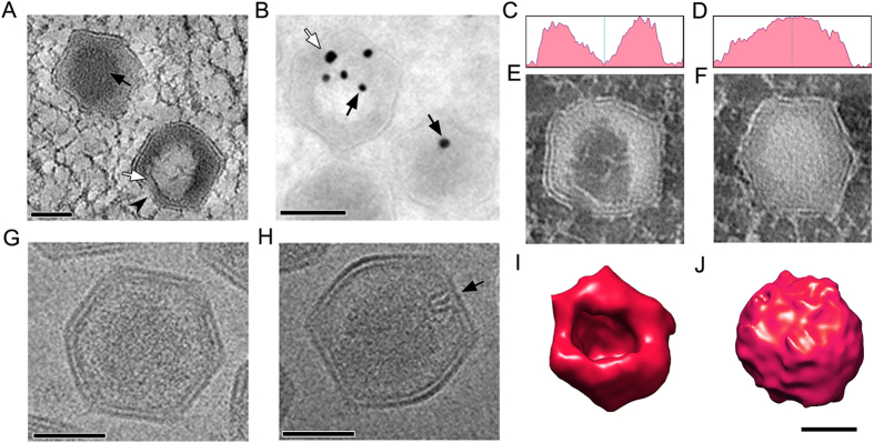
Figure 3. DNA encapsidation.
(A) A 220 nm tomographic slice of the VAS area revealed partially and fully formed viral particles. In mature virions, electron dense materials are packed to form a compact core inside the viral particle (black arrow), whereas immature virus have a small opening on one side (arrowhead). The electron dense fibrous material forms a lining on the viral capsid inner layer (white arrow); scale bar = 100 nm. (B) Immunogold labeling of Tokuyasu section of SGIV-infected cells with anti-viral DNA conjugated with 15 nm gold markers (black arrows) confirmed that the dense materials are viral DNA. Counter-labeling with anti-ORF075 (a DNA core associated protein) conjugated with 25 nm gold markers (white arrow) indicated that ORF075 is located next to the particle opening. Antibodies conjugated with gold can label loosely-packed DNA in the early stage of DNA encapsidation, but the affinity of gold conjugated antibody to label compacted DNA in mature virions is markedly reduced; scale bar = 100 nm. (C,D) Density plots of the central part of the immature and mature virions, respectively. (E,F) Inverted contrast imaging of the cross-sections of the corresponding viral particles in (C,D) demonstrates the density of the packaged genome in SGIV. (G,H) CryoEM of mature virions revealed the asymmetrical hairpin-shaped complex on one side of the capsid (black arrow). The hairpin structure can only be observed under EM when the sample is at a certain orientation. (I,J) 3D isosurface rendition of a partially formed particle (I) shows a clear opening in comparison with a fully formed, closed mature particle (J); scale bar = 100 nm.

