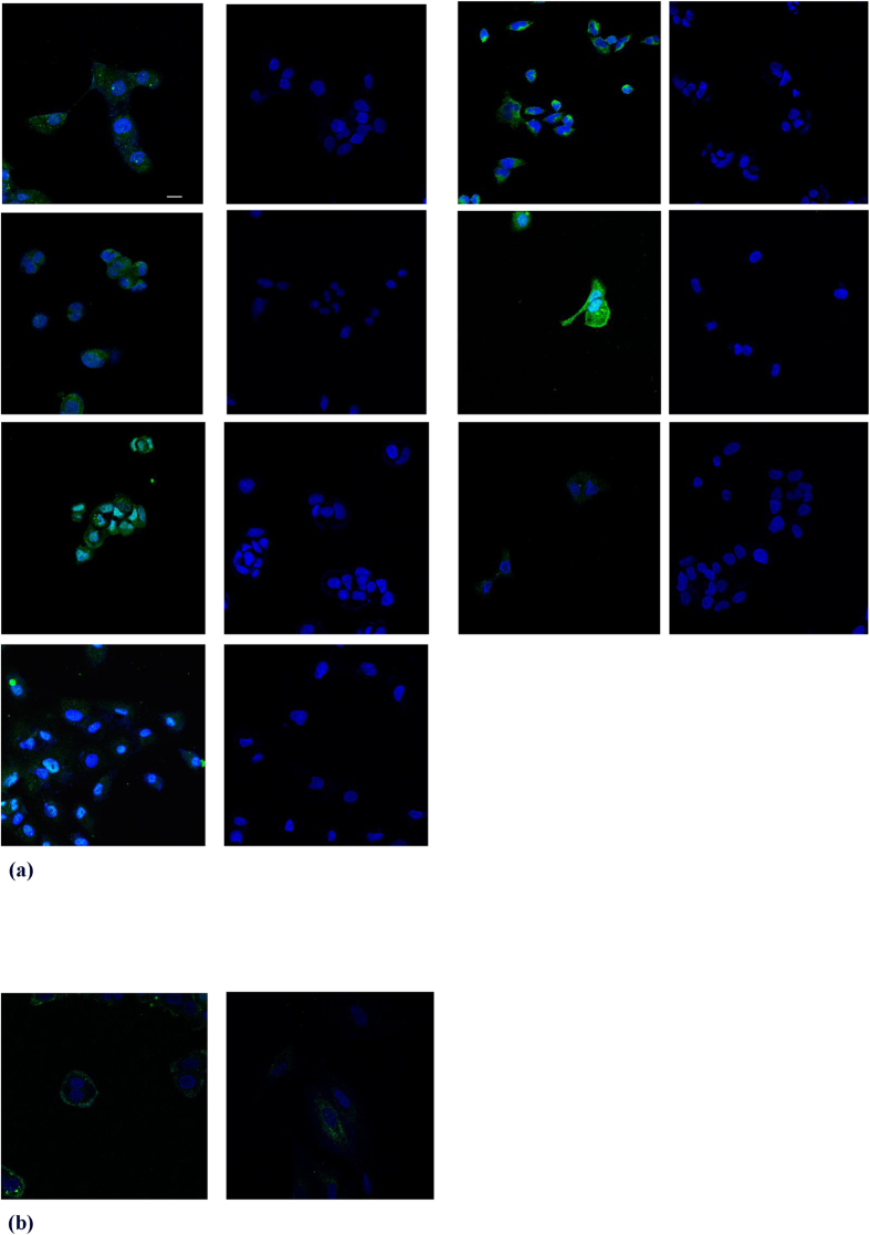
Figure 1. Representative confocal internal plane images of cell lines.
(a) confocal images of the internalization of the hexapeptide in the cytoplasm. In the first column respectively PaTu-8902, A2780, MCF-7, HuH-7 and in the second column the control of each adjacent cell line; in the third column respectively A375, DU145, MCF-10A and in the fourth column the control of each adjacent cell line. Note the green fluorescence dispersed in the cytoplasm. No labelling of the controls was observed. This figure is a merged image of green and blue fluorescence images (exc. 488 nm Argon Laser/em. BP500–550 filter). Scale bar = 14 μm for all images except for A375 and its control = 25 μm. (b) confocal images of the internalization of the scrambled hexapeptide in the cytoplasm of MCF-7 and MCF-10A cell lines. Scrambled does not enter both cells. It is detectable outside the cell membrane. Controls are not shown.

