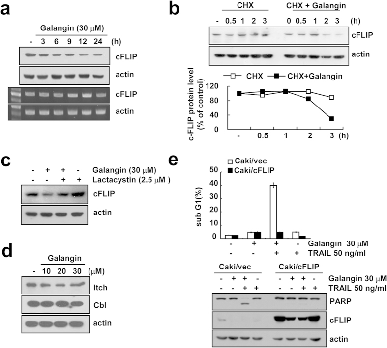
Figure 3. Galangin reduces protein level of cFLIP by post-translational modulation.
(a) Caki cells were treated with 30 μM galangin for the indicated time periods. Protein level and mRNA level of cFLIP were examined by western blotting and RT-PCR, respectively. Actin was used as a loading control in analysis. (cropped, full-length blots are in Supplementary Fig. S6). (b) Caki cells were pretreated with 20 μg/ml cycloheximide (CHX) for 30 min, and then treated with 30 μM galangin for the indicated time periods. Protein level of cFLIP was determined by western blotting. Actin was used as a loading control. The band intensity of cFLIP was measured using the public domain JAVA image-processing program ImageJ. (cropped, full-length blots are in Supplementary Fig. S6). (c) Caki cells were pretreated with 2.5 μM lactacystin for 30 min and then treated with 30 μM galangin for 24 h. Protein level of cFLIP was determined by western blotting. Actin was used as a loading control. (cropped, full-length blots are in Supplementary Fig. S6). (d) Caki cells were treated with indicated concentrations of galangin for 24 h. Protein levels of Itch and Cbl were determined by western blotting. Actin was used as a loading control. (cropped, full-length blots are in Supplementary Fig. S6). (e) Vector cells harboring empty vector (Caki/vec) and cFLIP overexpressing cells (Caki/cFLIP) were treated with 50 ng/ml TRAIL and 30 μM galangin for 24 h. The sub G1 population was measured by flow cytometry. The protein levels of PARP and cFLIP were determined by western blot analysis. Actin was used as a loading control. (cropped, full-length blots are in Supplementary Fig. S6).

