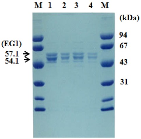
Figure 2.

SDS-PAGE of EG1. The gel was stained with Coomassie Blue R250. Endoglucanase (EG1) was identified using an MUC zymogram assay (data not shown). M, Protein molecular markers; lane 1 (10 μL) and lane 2 (2 μL) from fraction E1 and lane 3 (10 μL) and lane 4 (2 μL) from fraction E2 of the microcrystalline cellulose column eluent. SDS-PAGE, sodium dodecyl sulfate-polyacrylamide gel electrophoresis.
