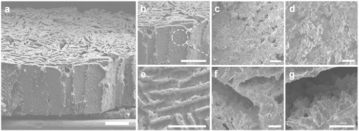
Figure 2. Freeze-cast Cu foam of macroporous layer-by-layer assembly.
(a) SEM image of a freeze-cast Cu foam showing a vertically aligned Cu multilayer assembly. Scale bar, 400 μm. (b–d) Side view SEM images of the macroporous layer-by-layer Cu foam. (b) An enlarged portion of the Cu foam. Scale bar, 400 μm. (c) A porous Cu layer resulted from dendritic growth of ice crystals. Scale bar, 40 μm. (d) The Cu layer with interconnected microscale pores is also confirmed at higher magnification. Scale bar, 40 μm. (e–g) Top view SEM images of the macroporous layer-by-layer Cu foam. (e) A randomly oriented layered structure. Scale bar, 400 μm. (f) The development of dendrites in side view simultaneously observed from (c,d) in front view. Scale bar, 40 μm. (g) The dendritic-like morphology character is also confirmed at higher magnification. Scale bar, 40 μm.

