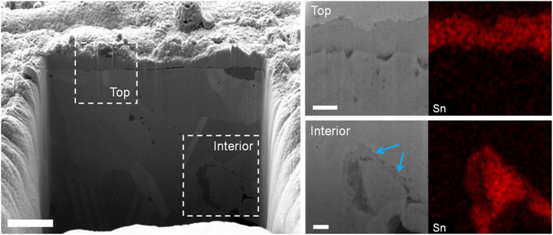
Figure 3. SnO2-coated Cu foam electrode.
Cross-sectional SEM images of a SnO2/Cu foam electrode in its entirety and at top and interior regions as indicated by the white dotted rectangles with Sn element mapping. The blue arrows indicate interior SnO2 coating layer surrounding a secondary pore. Scale bars, 2 μm (entirety), 500 nm (top) and 500 nm (interior), respectively.

