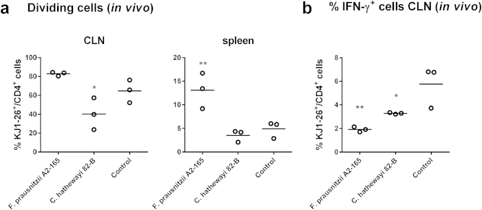
Figure 6. Percentage of dividing and IFN-γ+ OVA-T cells in vivo.
CFSE labelled naive OVA-T cells (KJ1-26+/CD4+) were adoptively transferred in BALB/c mice, after 24 h, mice were administered i.n. with bacteria plus OVA and after additional 72 h, OVA-T cells were isolated from cervical lymph nodes (CLNs) and spleens and analysed. (a) Percentage of dividing OVA-T cells (KJ1-26+/CD4+) isolated from CLNs or spleens. (b) Percentage of IFN-γ+ OVA-T cells (KJ1-26+/CD4+) isolated from CLNs. *indicates p < 0.05, **indicates p < 0.01 compared to the control administered OVA alone.

