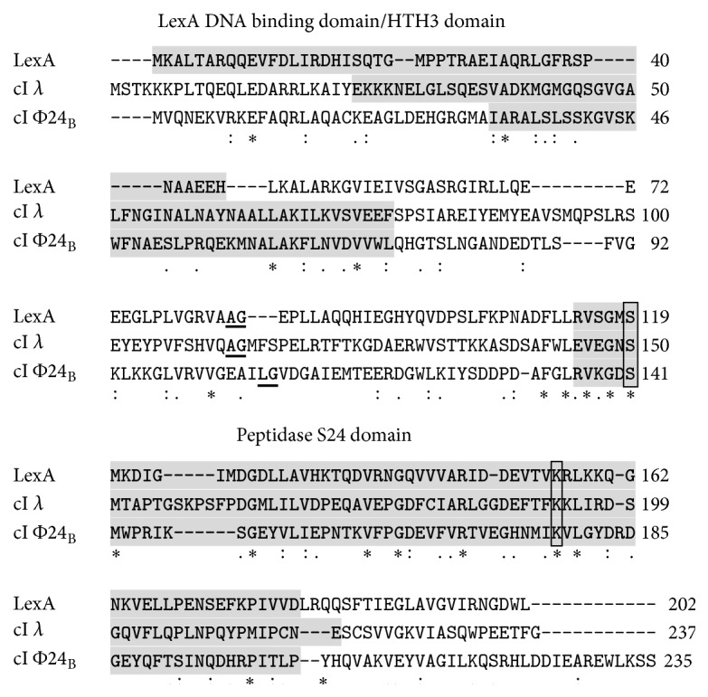
Figure 3.

Alignment of amino acid sequences of E. coli LexA protein and cI repressors of bacteriophages λ and Φ24B. Specific protein domains are indicated by grey background. Self-cleavage sites are underlined (two amino acid residues between which the cleavage occurs). The active sites of the peptidase domains are framed. Symbols under the sequence alignment indicate conserved sequence (∗), conservative mutations (:), semiconservative mutations (.), and nonconservative mutations ().
