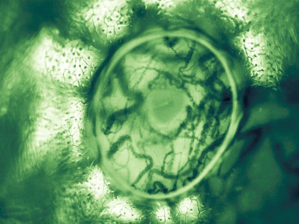
Figure 3.

Sublingual microcirculation image with a trapped bubble showing the microcosm of sublingual vascular and parenchymal cells. A bubble trapped under the lens cap and provided an optical effect whereby extra magnification and contrast was achieved at the perimeter of the image. Seen in the center is the opening to a submandibular duct surrounded by sublingual microcirculation consisting of red and white blood cells flowing in capillaries and venules.
