Figure 6.
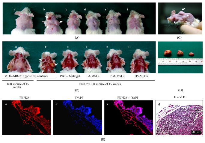
Tumor formation in mesenchymal stromal/stem cells (MSCs) transplanted into immunodeficient mice. (A) and (B) Images obtained after elimination of dorsal hair or clothing, respectively, in each mouse. (a–f) Cells (1 × 10⁷) stained with PKH26 were transplanted to both dorsal subcutaneous spaces of ICR mice (a) or NOD/SCID mice (b–f), and the mice were sacrificed after 15 weeks to confirm tumor formation. ((A)a-b) and ((B)a-b) show mice injected with MDA-MB231 as a positive control and c–f show mice injected with PBS + Matrigel (M), A-MSCs + M, BM-MSCs + M, and DS-MSCs + M, respectively. (C) and (D) display tumors isolated from mouse (A)b (Figures 6(A)-6(b)). ((E)a–c) Immunohistochemical staining and ((E)d) H&E staining were performed in tissue sections from tumors. ((E)a) and ((E)b) Red and blue identify the PKH26-stained membrane of cells and counterstaining of nucleic acids, respectively. Arrows indicate the blood vessels. Scale bars = 500 μm.

