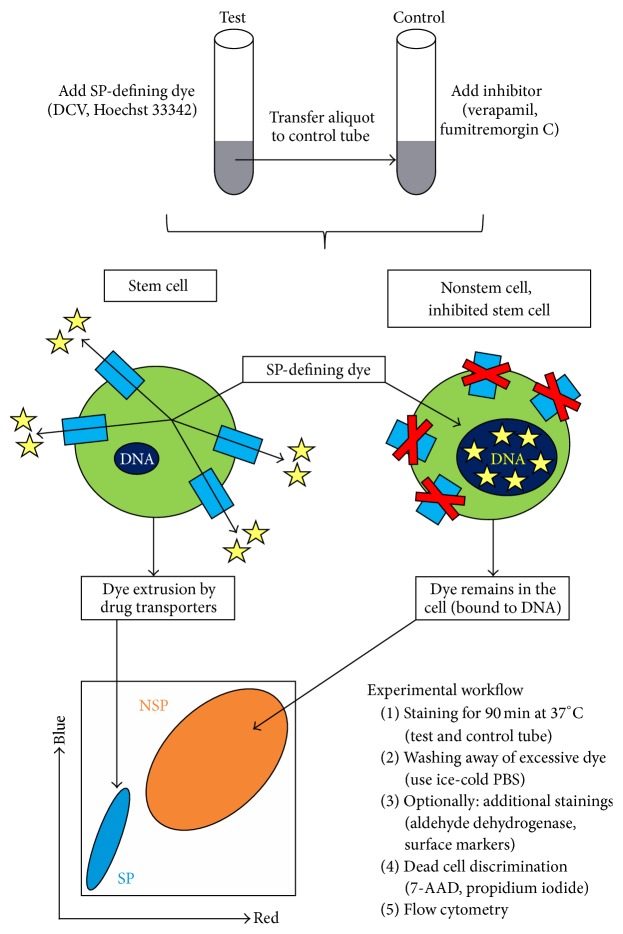
Figure 1.
Principle and workflow of SP detection. SP-defining dyes are lipophilic and enter cells passively to target nuclear and mitochondrial DNA. However, binding to DNA occurs only in drug transporter-deficient (nonstem) cells, whereas stem cells prevent this event through transporter-mediated dye extrusion, a process that is suppressed upon compound inhibition. The resulting differential dye accumulation enables flow cytometric discrimination of stem cell-enriched SP and a corresponding NSP containing the bulk of differentiated cells. The experimental workflow and the underlying principle are depicted. Dye molecules are shown as yellow asterisks and membrane-spanning ABC drug transporters are shown as light blue rectangles. Dark blue ellipses within the cells refer to DNA in both the nuclear and mitochondrial compartment.

