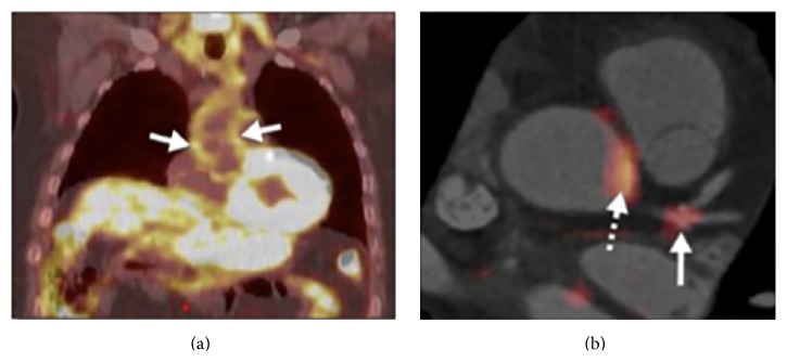
Figure 3.
Imaging arterial inflammation using (a) FDG-PET patient demonstrating enhanced aortic uptake of FDG on PET scan, indicating inflammation in the arterial wall due to atherosclerosis. (b) Coregistered FDG-PET/computed tomography images showing FDG uptake at the left main coronary artery trifurcation (solid arrow) in a patient with acute coronary syndrome. Aortic FDG uptake is indicated by the dashed arrow. In such patients, both aortic and coronary artery FDG uptake was increased compared with patients with stable coronary artery disease. Reprinted with permission from Rudd et al. [47].

