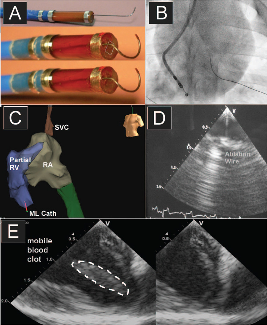
Fig. 3.
The MicroLinear (ML) catheter is shown in panel A with a small RF ablation wire integrated into the device at the tip but under full steering control by the operator. Panels B and C are the fluoroscopic and NavX mapping images, respectively, both showing the MicroLinear catheter near the RV apex in the pig. Panel D shows the clear delineation of the RF ablation wire, and panel E demonstrates the high level of image quality of this small 24-element phased-array forward-looking catheter.

