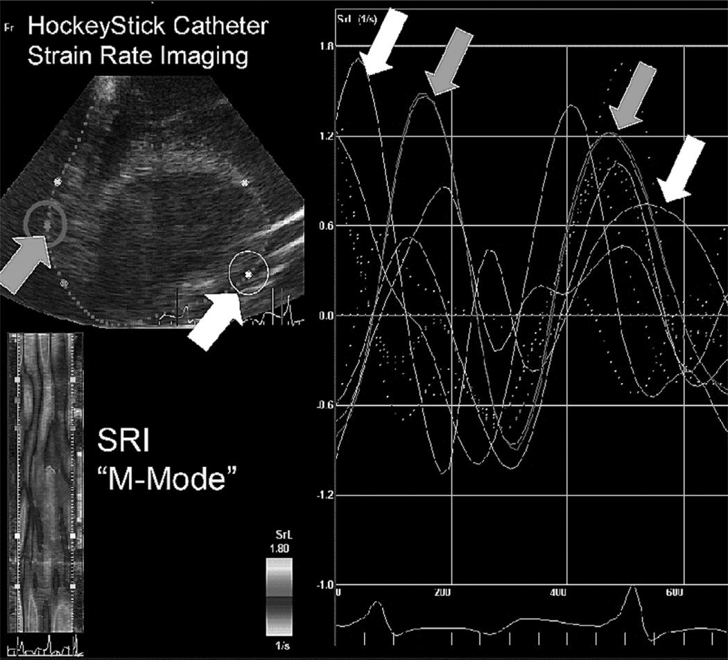
Fig. 6.
A HockeyStick catheter used for intracardiac strain rate imaging while open chest pacing is conducted in the pig. The image frame at upper left shows 5 SRI tissue “target points” at various LV wall positions in the short axis view from the RA. Two of the wall target points (white and gray arrows at left), tracked according to their 2-D strain rate time plot at right, show a loss in phasic synchrony as a result of epicardial pacing electrode stimulation. The plot limits are −1.0 to 1.8 s−1 in strain rate, and 0 to 700 msec in time duration. The pig heart rate is approximately 150 min−1.

