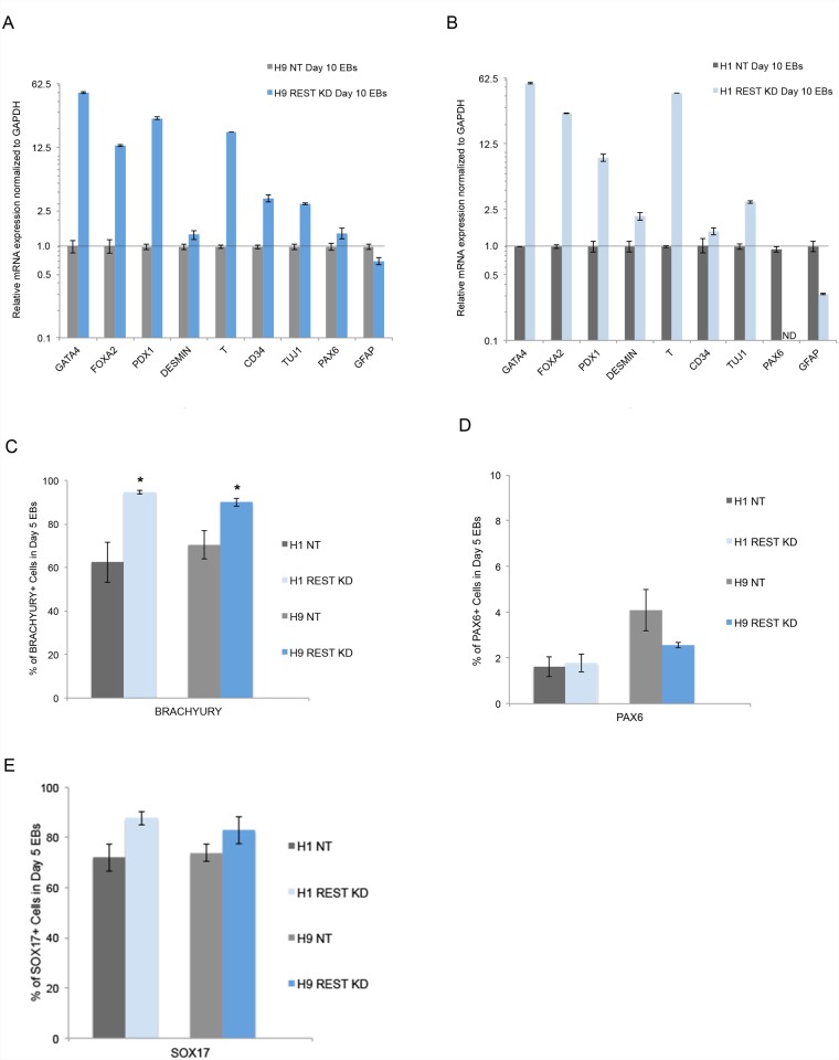
Fig 3. REST KD cells have increased mesendoderm lineage differentiation bias in vitro.
A-B. Evaluation of in vitro differentiation potential in Day 10 embryoid bodies (EBs). QPCR analysis revealed an increase in expression of endoderm and/or mesoderm markers in both H9 (A.) and H1 (B.) REST KD day 10 EBs. Shown are representative graphs of lineage marker analysis for each of the three germ layers. Error bars represent standard error of the mean (SEM) from three technical replicates. ND = Not detected. C-E. Evaluation of protein changes using FACS analysis across two independent REST KD lines (H9, H1) revealed an increase in expression of the mesendoderm marker BRACHYURY compared to control NT lines but no change in the ectoderm marker PAX6 or endoderm marker SOX17. Error bars represent SEM of three independent experiments and asterisks denote a p value < .05 using Student’s t-test analysis.

