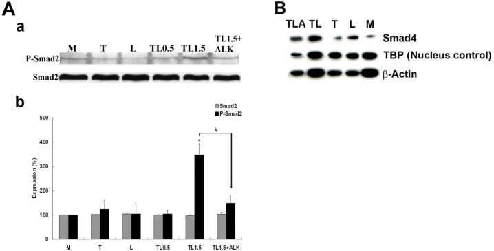
Fig 2. Western Blots of Smad2 phosphorylation at 1.5 h and nuclear expression of Smad4 at 3 h.
A) Smad2 and Phospho-Smad2 protein. Western Blot (a) and densitometry quantification (b) of Lysates from endothelial cells treated with TGRL (150 mg/dL) + LpL (2 U/mL) TL for 1.5 h (TL1.5) show significantly increased phosphorylation of Smad2 compared to cells treated with media (M), TGRL (T), LpL (L) or TL for 0.5 h (TL0.5). Addition of 10 μM of ALK, TGF-β receptor inhibitor, effectively blocks Smad2 phosphorylation (TL1.5+ALK). N = 3/treatment group, P≤0.05 as significant, * = TL1.5 compared to M, L, T, or TL0.5, # = TL1.5+ALK compared to TL1.5. B) Smad4 nuclear expression comparing M, L, TL and TLA. Cytosolic fractions were also run on the same blot corresponding to the nuclear fraction. Levels of Smad4 were too low to detect when loaded at a proteins concentration equivalent to the level of protein loaded for the nuclear fraction. These lanes were removed for clarity. Addition of 10 μM of ALK, TGF-β receptor inhibitor, suppressed but did not completely abrogate lipolysis induced Smad4 nuclear accumulation.

