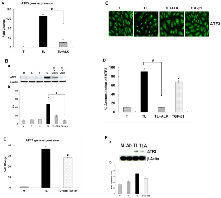
Fig 6. The effect of the ALK 4, 5 and 7 inhibitor or anti TGF-β antibody on the TGRL lipolysis induced ATF3 expression.
HAEC were exposed to TGRL (T), TGRL lipolysis products (TL) or 20 ng/mL human TGF-β1 for 3 h. TGF-β receptor inhibitor, ALK significantly suppressed: A) mRNA expression of ATF3. N = 3, P≤0.05. * = TL compare to T, # = TL with 10 μM of inhibitor ALK (TL+ALK) compared to TL. B) Western blot (a) and densitometry quantification (b) for ATF3 protein. N = 3, P≤0.05. * = TL compare to T, # = TL+ALK compared to TL. TL+CD36 antibody as control (positive/negative). C) Immunofluorescence images showing nucleus accumulation of ATF3. N = 3 coverslips/treatment group, Bar = 20 μm. D) % Translocation of ATF3. N = 6 coverslips/treatment group, P≤0.05, * = TL or TGF-β1 compare to T, # = TL+ALK compare to TL, Bar = 20 μm. anti TGF-β1 antibody (Ab) suppressed: E) mRNA expression of ATF3 was significantly suppressed. N = 3, P≤0.05. * = TL compare to M, # = TL+ anti TGF-β1 antibody compared to TL. F) Western blot (a) and densitometry quantification (b) ATF3 protein expression was trend toward suppressed significant. N = 3, P≤0.05. * = TL or TL + anti TGF-β1 (TLA) compare to M. Ab = anti TGF-β1 antibody.

