Figure 2.
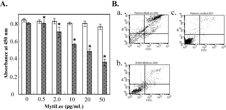
Reduction of cell viability by MytiLec. (A) Determination of viability by WST-8 assay. Dotted columns: Ramos. White columns: K562. Cells (2 × 10⁵ of Ramos; 5 × 10⁵ of K562) were incubated with various MytiLec concentrations as shown. Error bars: SE (n = 3); (B) Annexin V-binding and propidium iodide (PI) incorporation in MytiLec-treated cells. Horizontal axis: binding of FITC-labeled annexin V. Phosphatidylserine externalization and PI incorporation were evaluated by FACS analysis using MEBCYTO apoptosis kit. Ramos (a,c) and K562 (b) cells were treated with MytiLec (a,b: 20 μg/mL; c: 0 μg/mL) for 30 min at 4 °C. Data shown are mean values with error bars = SD of triplicate experiments. Asterisks = significant differences (p < 0.05) between treated and control groups.

