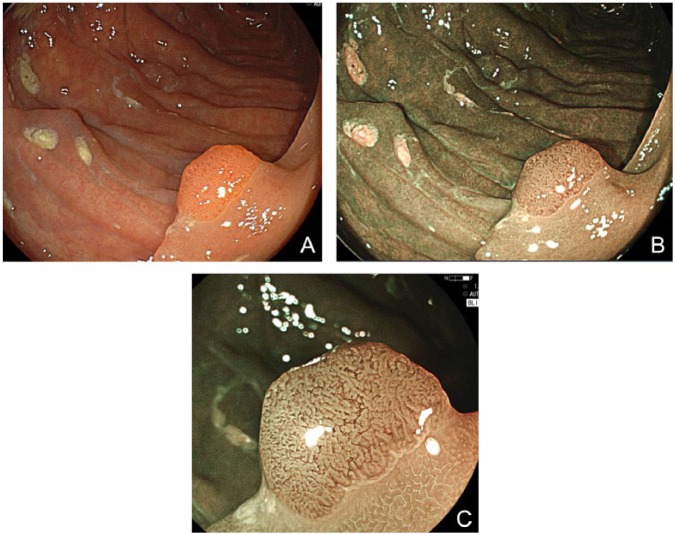
Figure 3.
(A) White-light mode. A sessile adenoma measuring 6 mm in size is clearly identified, but its superficial structure is not depicted clearly. (B) BLI-bright mode. Surrounding normal mucosa is depicted clearly and brightly even in a distant view. The vessel pattern of the polyp as well as the surface pattern is clearly depicted without magnification. (C) BLI mode. Magnified view shows a regular structure, suggesting a low-grade adenoma.

