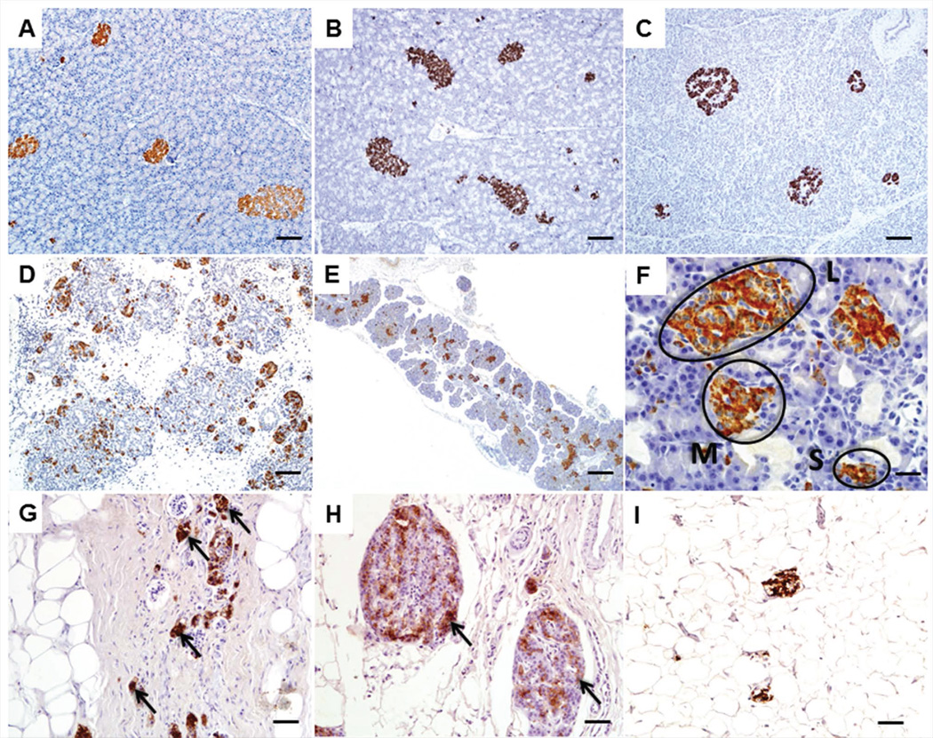
Figure 2.
Insulin immunohistochemistry (IHC) images of pancreatic islets from CF pigs, ferrets and human patients. A, B, C. Adult pancreata stained with insulin from WT pig (A), WT ferret (B) and normal human (C) Bars = 100 µm. D. Newborn CF pig pancreas stained with insulin to show abundance of islets even with exocrine pancreatic destruction. Bar = 200 µm. E. Insulin IHC of newborn CF ferret pancreas. Bar = 200 µm. F. Higher magnification images of B demonstrating the different sized islets present. There were more small islets (S) in CF ferrets compared to WT with fewer large (L) islets while medium (M) sized islets were not different (19). Bar = 20µm. G, H. Insulin IHC performed on human CF patients highlighting the lack of exocrine tissue with remnant insulin immunoreactive islets (arrows). I. Insulin IHC on a human CF patient with CFRD demonstrating a paucity of insulin immunoreactivity and abundance of adipose tissue.

