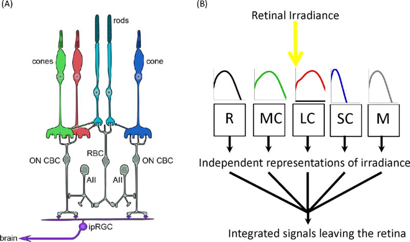
Figure 1. All retinal photoreceptor classes are upstream of circadian, neuroendocrine and neurobehavioral responses to light.

A. Schematic of the relevant retinal circuitry in humans. Non-image-forming responses originate in the retina and have been attributed to a particular class of retinal ganglion cell (ipRGC). ipRGCs are directly photosensitive owing to expression of melanopsin, which allows them to respond to light even when isolated from the rest of the retina. In situ they are connected to the outer retinal rod and cone photoreceptors via the conventional retinal circuitry. The details of their intraretinal connections are incompletely understood and probably vary between different subtypes. Shown here are major connections with on cone bipolar cells (on CBCs) connecting them to cone and, via amacrine cells (AII) and rod bipolar cells (RBC), rod photoreceptors. As a consequence, the firing pattern of ipRGCs can be influenced both by intrinsic melanopsin photoreception, and extrinsic signals originating in rods and each of the spectrally distinct cone classes (shown in red, green and blue). B. This feature is conceptualized in much simplified form, as a number of photoreceptive mechanisms (depicted as R for rod opsin; M for melanopsin; SC for S cone opsin; MC for M cone opsin; and LC for L-cone opsin), each of which absorbs light according to its own spectral sensitivity profile (shown in cartoon form as plots of log sensitivity against wavelength from 400 to 700 nm) to generate a distinct measure of illuminance. These five input signals are then combined by the retinal wiring, and within the ipRGC itself, to produce an integrated signal that is sent to non-image-forming centers in the brain. As each of the five representations of weighted irradiance is produced by a photopigment with its own spectral sensitivity profile, their relative significance for the integrated output defines the wavelength dependence of this signal, and hence of downstream responses.
