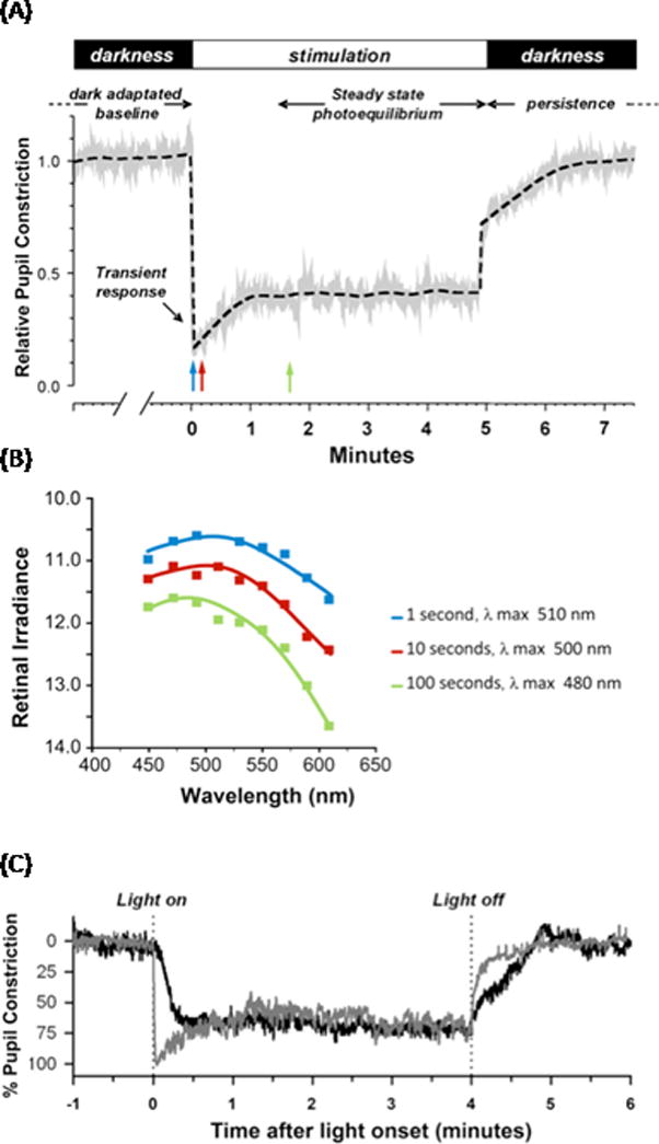
Figure 2. Spectral sensitivity of half-maximal pupillary constriction in humans.

(A). The pupillary light reflex response is composed of several different temporal components. At light onset, the pupil shows a rapid, transient constriction during the first 1000 ms of light exposure. This is followed by redilation to a tonic or sustained pupil diameter that stabilizes to a steady state constriction (photoequilibrium) even during prolonged constant illumination. When the light is turned off, there is a slow, delayed redilation of the pupil back to the resting (dark-adapted) state that is melanopsin-driven. Graph adapted from [76]. B. Mean spectral sensitivity is depicted as the retinal irradiance (log quanta/cm2/s) required to elicit a criterion pupil response (half maximal constriction) at nine wavelengths for three different stimulus durations of 1, 10, and 100 s (corresponding to the positions of blue, red, and green arrows in A). The smooth curve through the data points represents the optimal fit to the data using a mathematical combination of rod, cone, and melanopsin spectral sensitivities. As the stimulus duration increases, the sensitivity of the response gradually decreases by more than one log unit and is shifted towards shorter wavelength, from 510 nm at 1 s, to 500 nm at 10 s, and 480 nm at 100 s. Graph derived from data in [77]. (C) Representative traces for pupillary constriction in a sighted participant (gray) and in a blind individual without rod and cone function (black). Pupillary constriction to 480 nm was sluggish and lacks the transient response after light onset in the blind individual, whereas the sustained steady state pupillary light reflex (PLR) and the persistent response when the light is turned off are conserved. Graph reproduced, with permission, from [29].
