Figure 1.
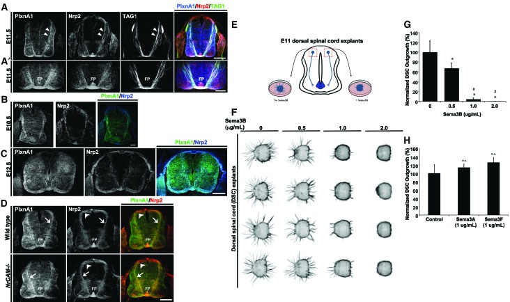
Dorsal commissural precrossing neurons express Nrp2 and PlxnA1, and their axons are responsive to Sema3B inhibition in a dosage-dependent manner. (A) Representative confocal images of a transverse wild-type mouse spinal cord section at E11.5 labeled with antibodies against PlxnA1 (blue) (Yoshida et al. 2006), Nrp2 (red), and TAG1 (green). White arrowheads illustrate the coexpression of PlxnA1, Nrp2, and TAG1 on precrossing axons in the dorsal spinal cord. Bar, 125 µm. (A′) Higher-magnification images of the same spinal cord section shown in A illustrate colabeling of PlxnA1, Nrp2, and TAG1 in the ventral spinal cord and the ventral commissure. (FP) Floor plate. Bar, 75 µm. PlxnA1 antibody specificity is shown in Supplemental Figure 1. (B) Wild-type E10.5 spinal cord transverse section double labeled with antibodies against mouse PlxnA1 (left panel) and Nrp2 (middle panel) and merged (right panel). Bar, 60 µm. (C) Wild-type E12.5 spinal cord transverse section double labeled with antibodies against mouse PlxnA1 (left panel) and Nrp2 (middle panel) and merged (right panel). Bar, 250 µm. (D) E11.5 transverse spinal cord sections taken from wild-type and NrCAM-null animals and processed for immunolabeling against PlxnA1 and Nrp2. Dorsal commissural neuron cell bodies and their precrossing axons are labeled with white arrowheads and white arrows, respectively. Bar, 125 µm. (E) Schematic diagram illustrating the microdissection of E11 dorsal spinal cord explants for culture and analyzing precrossing axon outgrowth. (F) Representative dorsal spinal cord explant cultures challenged with increasing levels of Sema3B proteins. (G) Axonal outgrowth from dorsal spinal explants were normalized and are shown in percentages with respect to the untreated (100 ± 11.45) cultures. A decrease of >30% (67.54 ± 5.29), 95% (4.97 ± 1.22), and 100% (0.63 ± 0.02) in outgrowth was observed with 0.5 μg/mL, 1.0 μg/mL, and 2.0 μg/mL Sema3B, respectively. (H) Normalized axonal outgrowth of dorsal commissural precrossing axons treated with Sema3A and Sema3F at 1 μg/mL concentration. The graph shows percentages normalized to untreated controls (100 ± 10.55), Sema3A (113.6 ± 4.12), and Sema3F (125.6 ± 6.29). Data represent compiled means ± SEM from n = 4 independent explant cultures. Analysis of variance (ANOVA) followed by post-hoc Tukey test, (*) P < 0.05 compared with 0 μg/mL; (#) P < 0.05 compared with 0.5 μg/mL of Sema3B; (n.s.) not significant.

