Figure 2.
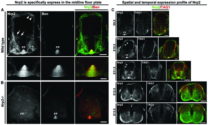
Nrp2 is highly expressed in the floor plate of the mouse spinal cord and is dynamically regulated during commissural axon pathfinding. (A) Wild-type E11.5 mouse spinal cord transverse sections show Nrp2 expression (green) in dorsal (white arrowheads) and ventral (white arrows) commissural neuron populations. Nrp2 is highly expressed in the floor plate and was colabeled with the floor plate-specific murine SC1-related protein (Ben; red). The bottom rows are a higher magnification of the top row images of the floor plate region. Bars: top row, 125 µm; bottom row, 25 µm. (B) A Nrp2 homozygote-null mutant (Nrp2−/−) spinal cord section at E11.5 colabeled with Nrp2 (green) and Ben (red). Bar, 125 µm. (C) Wild-type transverse spinal cord sections are shown colabeled with antibodies against Nrp2 (green) and TAG1 (red). Expression of Nrp2 in the floor plate can be detected as early as E9.5, at a developmental stage before commissural neurons extend axons toward the floor plate at the ventral midline. Bar, 100 µm. At E10.5, wild-type Nrp2-positive (green) commissural axons coexpressing TAG1 (red) have reached the ventral midline, where the floor plate robustly expresses Nrp2. Bar, 100 µm. At this stage, expression of PlxnA1 can also be detected in commissural axons, as shown in Figure 1C. Immunofluorescence of Nrp2 expression from both the midline crossing axons in ventral commissure and the floor plate cells is the highest at E11.5. Bar, 160 µm. While a few precrossing axons still express Nrp2 at E12.5, the floor plate-derived Nrp2 expression is down-regulated. Bar, 200 µm. By E13.5, the majority of commissural axons have crossed the midline, and the expression of Nrp2 from the floor plate has dramatically decreased in fluorescent intensity. Bar, 250 µm. White arrowheads point to dorsal commissural axons. (FP) Floor plate.

