Figure 1.
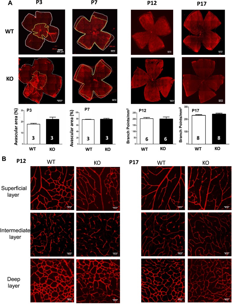
Genetic inactivation of A1Rs does not affect the normal development of retinal vessels in mice. (A) The development of retina vasculature of WT and A1R KO mice at P3, P7, P12, and P17 in room air was visualized by isolectin B4 staining in whole-mount retinas. Vascularized areas and whole retinal surface are shown by yellow dotted line and white dotted line, respectively. The superficial vascularized areas of retinas at P3 (n = 3/group) and P7 (n = 3/group) were quantified as a percentage of the whole retinal area. At P12 (n = 6/group) and P17 (n = 8/group), “branch point” analysis was used for quantification of retinal vascularization under room air. Data are presented as mean ± SEM. Scale bar: 500 μm. (B) The distributions of three retinal vascular layers were displayed in distinct confocal planes. The retinal images from P12 and P17 of WT and A1R KO in room air were examined by isolectin B4 staining of whole-mount retinas. The development of morphology and distribution of superficial, intermediate, and deep plexuses were indistinguishable between WT and A1R KO mice. Scale bar: 50 μm.

