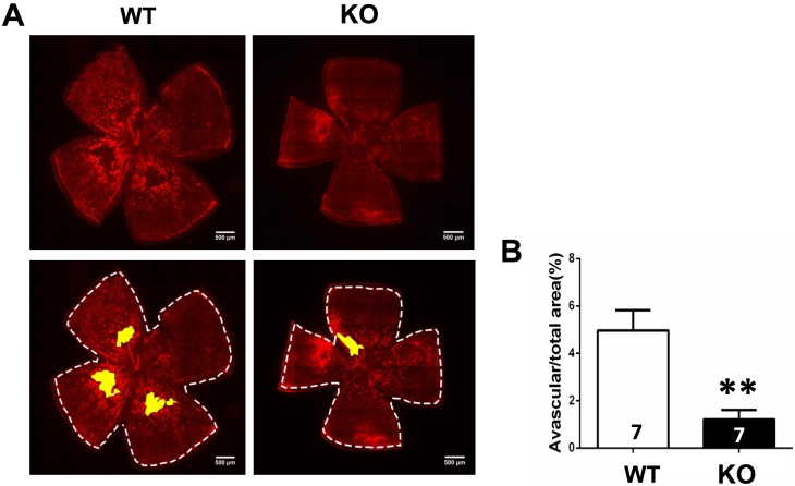
Figure 6.
Genetic deletion of A1Rs reduced retinal normal vascularization at P21 of OIR. (A) Retinal blood vessels of both WT and KO groups were visualized by isolectin B4 staining of whole-mount retinas at P21 of OIR. The whole retinal surface is shown by white dotted line. Avascular area is indicated as yellow. Scale bar: 500 μm. (B) Avascular area (%) was quantified as a percentage of the whole retinal surface (n = 7 retinas from seven mice for each group). Data are presented as the mean ± SEM. *P < 0.05, Student's t-test.

