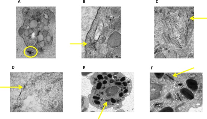
Figure 3.
Electron microscopic studies of four CHM patients' peripheral lymphocytes. Photos illustrate striking intracytoplasmic crystalline structures with their magnifications. (A) Patient 2 with 23,000× magnification, (B) patient 3 with 49,000×, (C) patient 3 with 68,000×, (D) patient 4 with 49,000×, (E) patient 7 with 8900×, and (F) patient 7 with 22,000×.

