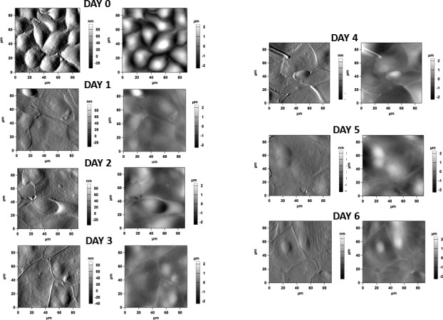
Figure 5.

Atomic force microscopy images of the cell cultures at day 0 (unstratified) and at days 1 to 6 during the stratification process. (Left) Figures represent phase images; (right) figures represent height images. Unstratified cells show cobblestone morphology, and are tall with no discernable cell junctions, resembling basal corneal epithelial cells. Once exposed to SM, cells flatten and develop marked cell junctions. No other noticeable topographic features were observed.
