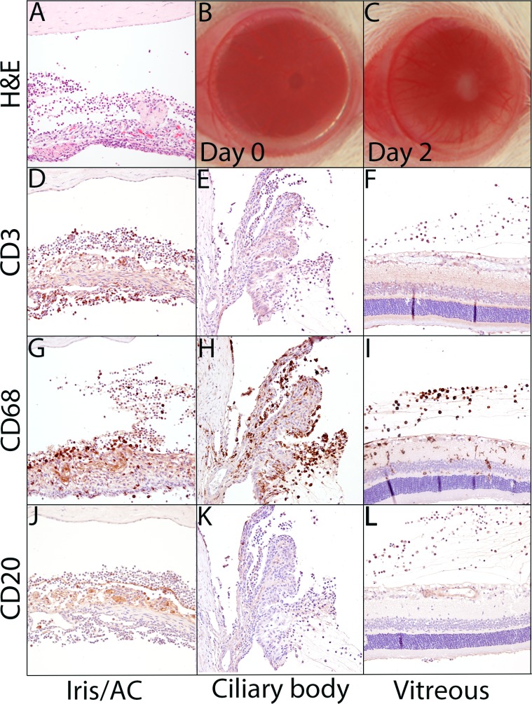
Figure 2.
Primed mycobacterial uveitis generates a CD68-predominant inflammatory response in the anterior chamber, vitreous, and surrounding the ciliary body without retinal degeneration. (A) Hematoxylin and eosin–stained section of iris and anterior chamber demonstrating inflammatory cells in the aqueous, iris vasculature dilation, and epithelioid macrophages forming a multinucleated giant cell on the anterior iris surface. (B) Uninflamed eye at PMU day 0, prior to intravitreal injection. (C) The PMU eye during peak inflammation (day 2) demonstrating dilated corneal vasculature, hypopyon, and pupillary membrane. Immunohistochemistry with anti-CD3 (D–F), anti-CD68 (G–I), and anti-CD20 (J–L) antibodies identifies a predominance of CD68- and CD3-positive inflammatory cells with few CD20-positive cells. The inflammation is predominantly noted in the anterior chamber (D, G, J), surrounding the ciliary processes (E, H, K), and in the vitreous (F, I, J). CD68-positive cells are seen within the retina (J), but retinal damage was not noted.

