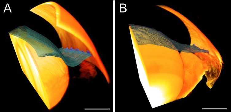
Figure 4.
Anterior chamber deepening in enucleated mouse eyes as visualized by 3D reconstructions of the anterior segment. Eyes perfused via the AC (A) show posterior iris displacement and an enlarged iridocorneal angle relative to eyes perfused via PC (B). Both eyes were perfusion-fixed at 40 mm Hg. The iris is shown in blue-green and the cornea and lens in yellow. Scale bars: 0.5 mm. Individual sections used to generate the 3D reconstructions are shown in Supplementary Fig. S5.

