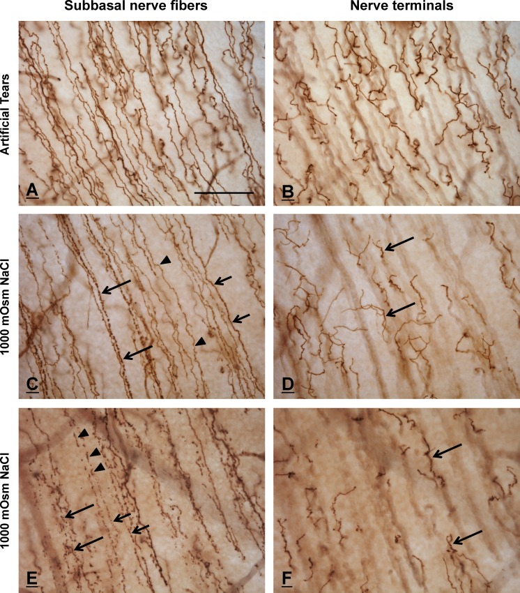
Figure 8.
Immunohistochemically stained SNFs and nerve terminals in AT-treated (A, B) and HOS-treated (C–F) corneas. Each of the paired images in rows 1 to 3 shows the same corneal area focused alternately on SNFs (left column) and nerve terminals (right column). (A, B) Subbasal nerve fibers (A) and nerve terminals (B) in a control, AT-treated cornea are smooth and morphologically intact. (C, D) Subbasal nerve fibers in a HOS-treated cornea (C) exhibit a variety of morphologies, ranging from smooth (arrowheads) to granular (short arrows) to prominently beaded (long arrows). In contrast, the nerve terminals that originate from these SNFs (arrows, [D]) appear structurally intact and are indistinguishable morphologically from those of control corneas. (E, F) Subbasal nerve fibers from a different area of the same HOS-treated cornea shown in (C) and (D), showing more profound degenerative changes. Most of the SNFs in this region have fragmented into linear arrays of disconnected axon fragments (arrowheads) or beads (short arrows). The SNFs at bottom left (large arrows) demonstrate a near-total loss of normal morphology, suggestive of an advanced stage of degeneration. Despite the profound changes in SNF morphology in this region, the nerve terminals (arrows, [F]) remain histologically intact. The regions of the subbasal plexi illustrated in the HOS corneas (C–F) are more peripheral than the area shown in the ATs cornea (A, B); thus, the nerve terminals, although morphologically intact, appear to be slightly less in number and density. The calibration bar in (A) is 50 μm and applies to all figures.

