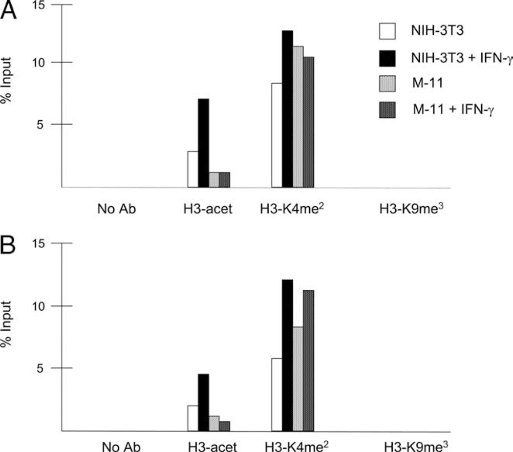
FIGURE 8.
Histone modifications at the IRF-1 and CIITA promoters in mouse fibroblasts and TBCs. NIH-3T3 and M-11 cells were cultured for 0 and 3 h with 500 U/ml IFN-γ, and subjected to ChIP assays using Abs specific for acetylated H3, acetylated H3-lysine 9 (K9), dimethylated H3-K4 (H3-K4me2), and trimethylated H3-K9 (H3-K9me3). Quantitative PCR was performed in duplicate for each sample using primers specific for the IRF-1 promoter (A) and CIITA pIV (B). Results for each histone modification are represented as the percentage of input, and are the average of at least two independent experiments.

