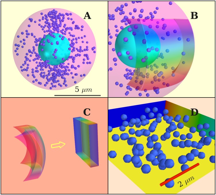
Fig 5.
(A) Simulated cell with diameter 10 μm. The diameter of vesicles is 300 nm. (B) Model for active zone, chosen as a thin layer close to cell membrane. (C) Map of the active zone onto a cuboid. (D) Cell volume in the simulations. Scale bar: 2 μm. Vesicles elastically bounce from all walls except from the membrane, where fusion is assumed to occur.

