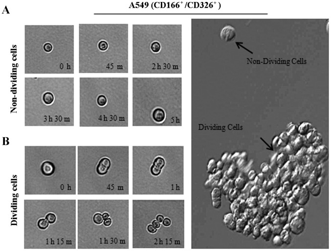
Figure 5.
Phase-contrast images of a single CD166+/EpCAM+ cell derived from A549 cells cultured in 96-well plates under anchorage-independent, serum-free conditions. Sorted CD166+/EpCAM+ cells either in the non-dividing phase (A) or actively dividing phase (B) were recorded at different time points as indicated.

