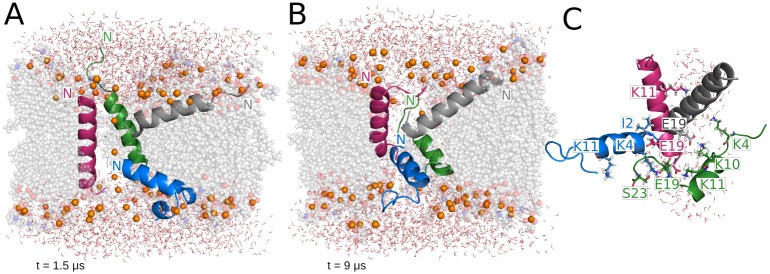
Fig 1. Representative structures from the MAG2 parallel tetramer simulation.
(A) Thin pore supported by an irregular distribution of MAG2 monomers. (B) Final structure. (C) Amino acids involved in intermolecular interactions formed during the last microsecond of the trajectory and pore waters. In all the figures, lipids are shown as grey spheres, phosphorus atoms representing the lipid headgroups are shown as orange spheres and water molecules as lines. Lipid spheres are semi-transparent and some of the molecules have been omitted for clarity.

