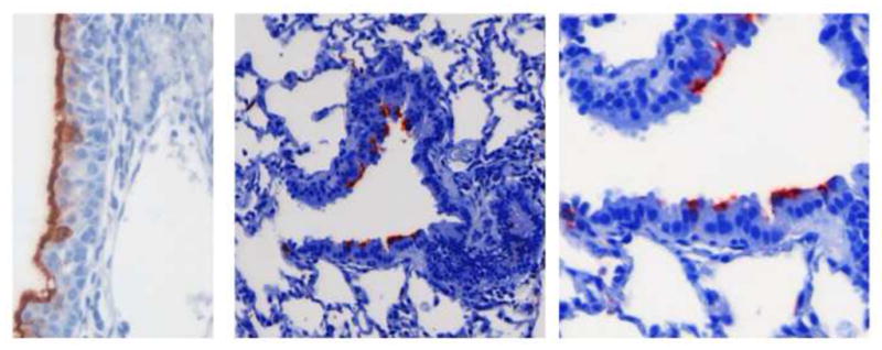
FIGURE 2.

In the cotton rat, the nasal cavity is susceptible to infection by hRSV, with diffuse immunostaining of the nasal mucosa (in red) as shown in the left hand panel. As with human patients, lower airway infection in the cotton rat involves the columnar epithelium lining the bronchioles, seen in the central and right hand panels. This is distinct from infection in the mouse model where it is primarily the alveolar lining cells that are infected by hRSV when lung sections are examined by immunohistochemistry.
