Fig. 1.

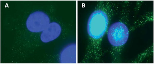
BRMS1 mainly distributed around the nucleus. Immunofluorescence images of (A) 435 and (B) 435/BRMS1 cells stained with anti-BRMS1 antibody (blue: nucleus; green: expression of BRMS1).

BRMS1 mainly distributed around the nucleus. Immunofluorescence images of (A) 435 and (B) 435/BRMS1 cells stained with anti-BRMS1 antibody (blue: nucleus; green: expression of BRMS1).