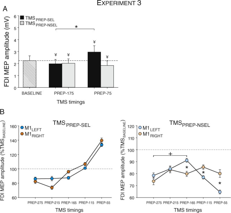
Fig. 8.

MEP amplitudes in Experiment 3. A: Evolution of MEPs elicited from a selected (MEPPREP-SEL) or non-selected (MEPPREP-NSEL) hand at TMSBASELINE and in pre-movement windows (TMSPREP-EARLY, TMSPREP-LATE). MEPs are suppressed at TMSPREP-175. At TMSPREP-LATE, excitability increased in the selected hand but remains suppressed in the non-selected hand. B: MEP amplitudes following M1RIGHT (filled dots) and M1LEFT stimulation (empty dots) for the different TMS epochs (see Methods section) in the selected (left) and non-selected hands (right). Note the absence of a HEMISPHERE effect for the selected handSEL. For the non-selected hand, inhibition in the left hand was relatively constant (M1RIGHT stimulation), but showed a non-monotonic profile in the right hand (M1LEFT stimulation). * and + = significantly different (p-value < 0.05). ¥ = significantly different (p-value < 0.05) from MEPs elicited at TMSBASELINE.
