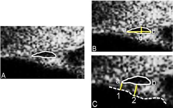
Fig 2. Example of Schlemm’s Canal and Trabecular Meshwork Measurements Made Using the iUltrasound Imaging System.

The black oval space shows Schelmm’s canal (SC). The meridional diameter of SC was measured from the anterior (a) to the posterior (b) end point of SC. To measure the coronal diameter of SC, we drew a vertical line across the canal to get two intersection points (c and d). The maximum distance between c and d was taken as the coronal diameter of SC. Lines 1, 2 indicated where trabecular meshwork thickness was measured and the dotted line shows the meshwork inner layer.
