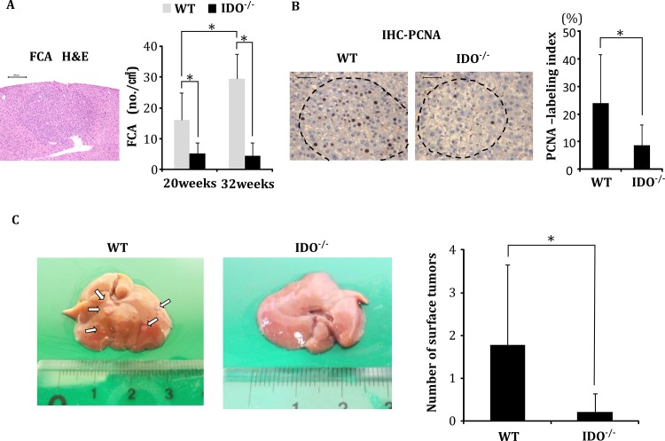
Fig 1. Analysis of pre-neoplastic lesions and nodules of DEN-treated mice.
Development of foci of cellular alteration (FCA), the proliferating cell nuclear antigen (PCNA)-labeling indices of the FCA, and the nodule frequency on the liver surface of DEN-treated experimental mice. (A) A representative photograph of a FCA that developed in DEN-treated WT mice at 32 weeks of age (H&E staining, left panel) and the average number of FCA in DEN-treated WT and IDO-/- mice at 20 and 32 weeks of age (right panel). (B) Representative photographs of PCNA-immunohistochemical analysis of the FCA that developed in DEN-treated WT mice and IDO-/- mice at 32 weeks of age (left panels). The PCNA-labeling indices of the FCA that developed in the experimental mice were determined by counting the number of PCNA-positive nuclei in the FCA (right panel). (C) Tumor nodules on the liver surface of DEN-treated WT and IDO-/- mice at 32 weeks. The white arrows indicate tumor nodules (left panel). The number of liver surface tumors in IDO-/- mice was significantly lower than that in wild-type mice (right panel). *, P<0.01 Data points represent the mean ± SD.

