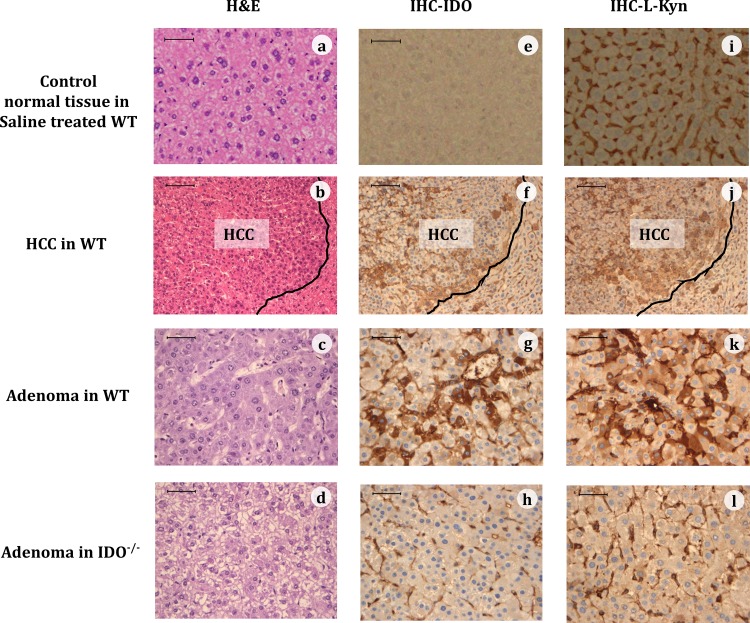
Fig 2. Representative immunohistochemical expression (IHC) of IDO and kynurenine (KYN).
Representative images of normal tissue (top panel) of a saline treated WT mouse, of HCC (second panels) and adenoma (third panels) of a WT mouse, and of adenoma of an IDO-/- mouse (bottom panels), at 32 weeks of DEN treatment that were stained with H&E (a-d), or were immunohistochemically stained for the IDO protein (e-h) or for L-KYN (i-l). The black line in the HCC tissues demarcates HCC from non-HCC tissue. (Bar = 50 μm)

