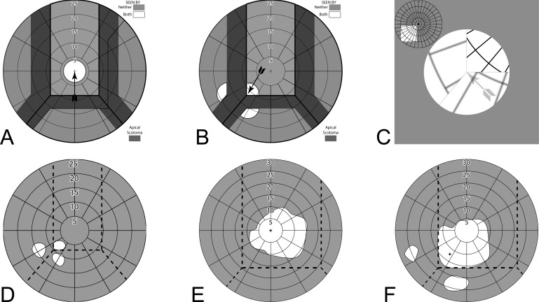
Figure 4.
InWave channel prism fields. (A) Calculated perimetry with 8° residual visual field at primary gaze in a 6-mm channel. The prisms have no effect. Field of view shaded darker is lost to the apical scotomas. (B) With gaze shifted 8.5° (of visual field) left and down to the channel corner (9° of diagonal ocular rotation), the field is split into three parts, giving a 6° extension left and 4° down, with corresponding gaps in between at the apical scotomas. (C) The percept diagram corresponding to (B) for this bilateral fitting shows the larger grid spacing of the shifted fields and perceived discontinuity (jump) in the field of view caused by the scotomas. Thin gray lines, not part of the patient's percept, are shown to identify the prism apices. There is no visual confusion or diplopia, nor field expansion. (D) Measured LE field of patient 1 at this gaze position (RE patched). The total area seen is slightly larger than the 8° residual field due to vignetting at the prism edges. Dashed lines indicate the apparent location of the prism apices, a bit to the right of the intended location. (E) The asymmetric monocular field of patient 2 required shifting the 12 mm (33°) channel to the right, so that the residual field at primary gaze lies entirely within the channel. (F) With fixation shifted 7° left and 9° degrees down, the channel corner splits the field. Even though the channel and residual field sizes differ greatly for these two patients, the extension provided is the same. The apparent difference in scotoma sizes is an artifact of the imprecision of these difficult measurements, also seen as the differences in field size and shape between (E) and (F) for the 20/500 acuity of this subject.

