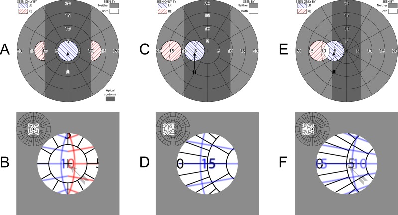
Figure 6.
Trifield prisms. (A) Simulated perimetry at primary gaze. The two 16Δ prisms add two peripheral islands of visibility. Two adjoining (but only monocular) apical scotomas, each larger than the patient's 8° field width, are evident (darker gray shading). (B) Corresponding percept diagram. Blue and red tints in the percept diagrams identify field viewed through the tinted right eye (RE) left (green) and right (red) prisms, respectively. (Although the prisms are tinted red and green, we diagram with red and blue in deference to dichromats.) Left eye (LE) maintains a direct, nonprismatic view at all gaze positions. True field expansion comes at the expense of central visual confusion. (C) With a left gaze shift of about half the residual field width, the RE view is entirely within the left green prism and a relatively continuous expanded view is afforded, without diplopia. (D) The corresponding percept diagram shows that there is still central confusion, but only from two views, not three. The digit 5 is coincidentally overlaid. (E) With 10Δ prisms, the power is not sufficient to avoid undesirable diplopia. (F) A region including the zero of the 10° eccentricity marking is thus perceived diplopically.

