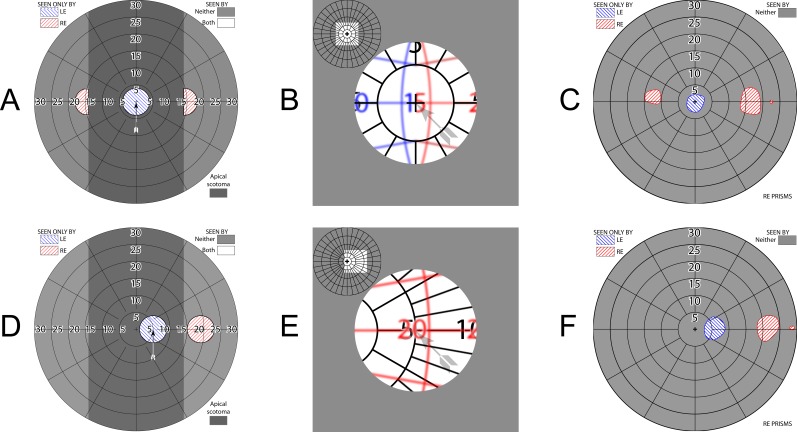
Figure 7.
Patient 1 Trifield results. The patient's fields have shrunk in the decade since the 25Δ prisms were fitted, resulting in larger field of view gaps than illustrated in Figure 6A (and the diagram scale has changed to accommodate the higher-power prisms). Unlike channel prisms, which provide no access to the scotoma areas even with gaze shifts, the fellow nonprism eye can see into the apical scotoma regions, so the loss (without head turning) is not as problematic. Having access at primary gaze to the region at eccentricities larger than the scotomas may be desirable, although that has not been studied. (A) Simulated perimetry at primary gaze. (B) Corresponding percept diagram shows the “Trifield” binocular central visual confusion. (C) Corresponding perimetry. The asymmetry is indicative of the difficulty of positioning the prism apices exactly at primary gaze, as the perimeter's fixation target is invisible at that location in the prisms, and the eyes readily dissociate into the phoria posture, without any clue for the directional shift needed to align the eyes to assess veridical direction. (However, since the prisms are intended to provide hazard detection followed by a gaze turn to foveate the hazard with the nonprism eye, precise angular perception may not be needed.) (D) With a 5° gaze shift toward the right, the right eye (RE) view is entirely through the right prism. (E) The corresponding percept diagram again illustrates that there is still central confusion showing, but only from two views, not three. (F) Corresponding perimetry. We use the symbology of our dichoptic perimeter, but dichoptic shutter goggles were not needed to identify each eye's obvious and separate contribution.

