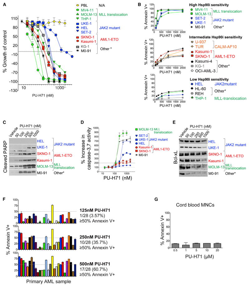
Figure 1. PU-H71 Is Cytotoxic in a Subset of AML Cells while Leaving Normal Blood Cells Unharmed.
(A) Growth of cells treated for 72 hr with PU-H71 (relative to untreated cells). PBL, peripheral blood lymphocytes.
(B) Cells were treated with or were not treated (control) with the Hsp90 inhibitor (Hsp90i), PU-H71. Percent annexin V+ cells relative to control after 48 hr treatment with PU-H71 is shown.
(C) Immunoblots showing cleaved PARP after 24 hr treatment with PU-H71.
(D) Percent increase in caspase-3, 7 activity assay after 24 hr treatment with PU-H71
(E) Immunoblots for Bcl-xL after 24 hr treatment with PU-H71
(F and G) Percent annexin V+ cells relative to control for primary AML cells (F) or cord blood mononuclear cells (MNCs) (G) after 48 hr treatment with PU-H71. (A, B, and D) Values denote mean ± SD (n = 4).
(F and G) Values denote SEM. See also Figure S1 and Tables S1 and S2.

