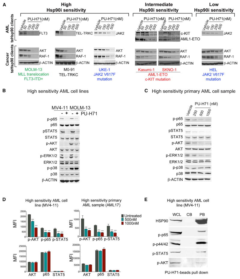
Figure 2. PU-H71 Treatment Depletes AML Cells of Major Leukemogenesis-Driving Proteins Irrespective of Their Apoptotic Sensitivity.
(A–C) Immunoblot analysis for (A) AML (FLT3, TEL-TRKC, JAK2, c-KIT, and AML-ETO) and cancer (AKT and RAF-1) teHsp90 clients after 24 hr treatment with PU-H71; (B and C) phosphorylated or total NF-κB p65, STAT5, AKT, ERK1/2, and p38 for leukemia cells treated for 6 hr with 1 μM PU-H71 are shown.
(D) Protein expression levels indicated as mean fluorescence intensity (MFI) for phosphorylated (upper panel) or total (lower panel) AKT, NF-κB p65, and STAT5. Values denote mean ± SEM. ***p < 0.001.
(E) Pull-down assay for MV4-11 cells using PU-beads followed by immunoblotting for the indicated proteins. CB, control beads; PB, PU-H71-beads; WCL, whole cell lysate.

