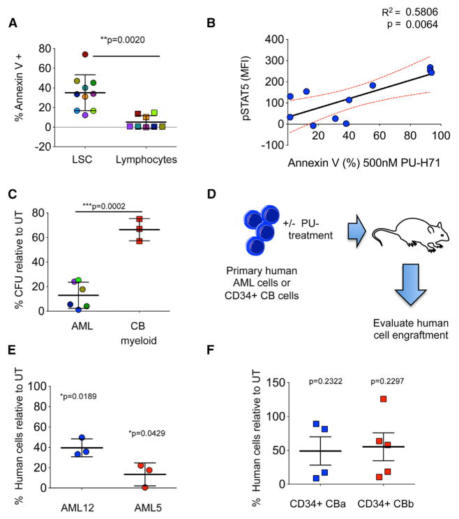
Figure 5. Dependence of AML Signaling Networks on teHsp90 Is Conserved at the Leukemic Progenitor and Stem Cell Level.
(A) Percent annexin-V-positive leukemia stem cells (LSCs) and lymphocytes in primary AML specimens treated with 500 nM PU-H71 for 48 hr. Each color represents an individual sample.
(B) Correlation between p-STAT5 (y axis) and sensitivity of LSCs to PU-H71 (x axis).
(C) Percent colony-forming units (CFUs) relative to untreated control (UT) for primary AML cells or CD34+ cord blood (CB) after 48 hr treatment with 500 nM PU-H71. Myeloid colonies are shown for normal CB cells.
(D) Schematic representation for the ex vivo engraftment assay to evaluate the effect of PU-H71 treatment on the ability of LSCs to initiate disease in immunodeficient mice.
(E and F) Percent engraftment of human AML cells (E) and CD34+ CB cells from two units (CBa and CBb) (F) after ex vivo treatment with PU-H71 for 24 hr. Each symbol represents an individual mouse.
Values denote mean ± SEM.

