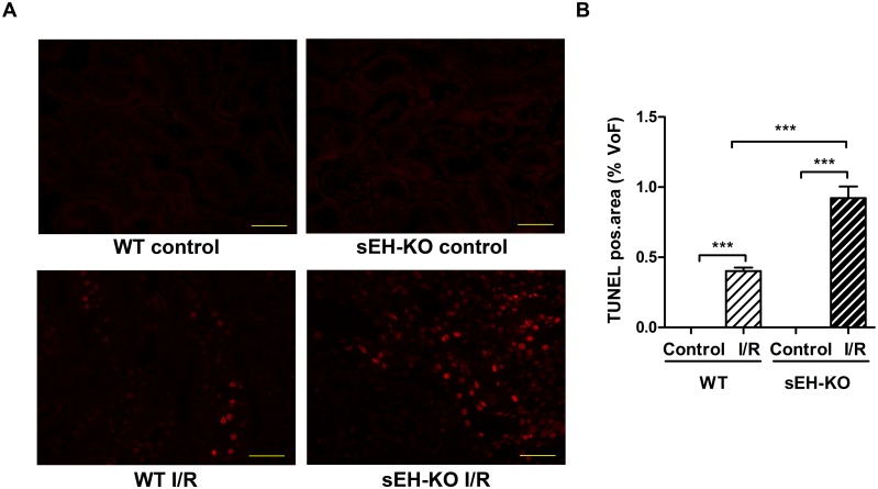
Fig 4. sEH gene disruption increased I/R-induced apoptosis of tubular epithelial cells.
(A): Representative images of renal sections after TUNEL-staining (magnification 400×, Scale bar 100 μm). Apoptosis was detected in the kidneys of all mice subjected to I/R-injury but not in the corresponding control mice. (B): Quantification of apoptosis in the cortex and outer medulla of kidneys harvested two days after reperfusion. The intensity of positively stained nuclei was related to the area of each chosen field of view (FoV) in the renal sections. sEH-KO mice displayed significantly stronger apoptosis compared to the WT-I/R or control groups. Data are given as mean ± SEM (n = 5 per group). ***p<0.001 vs WT.

