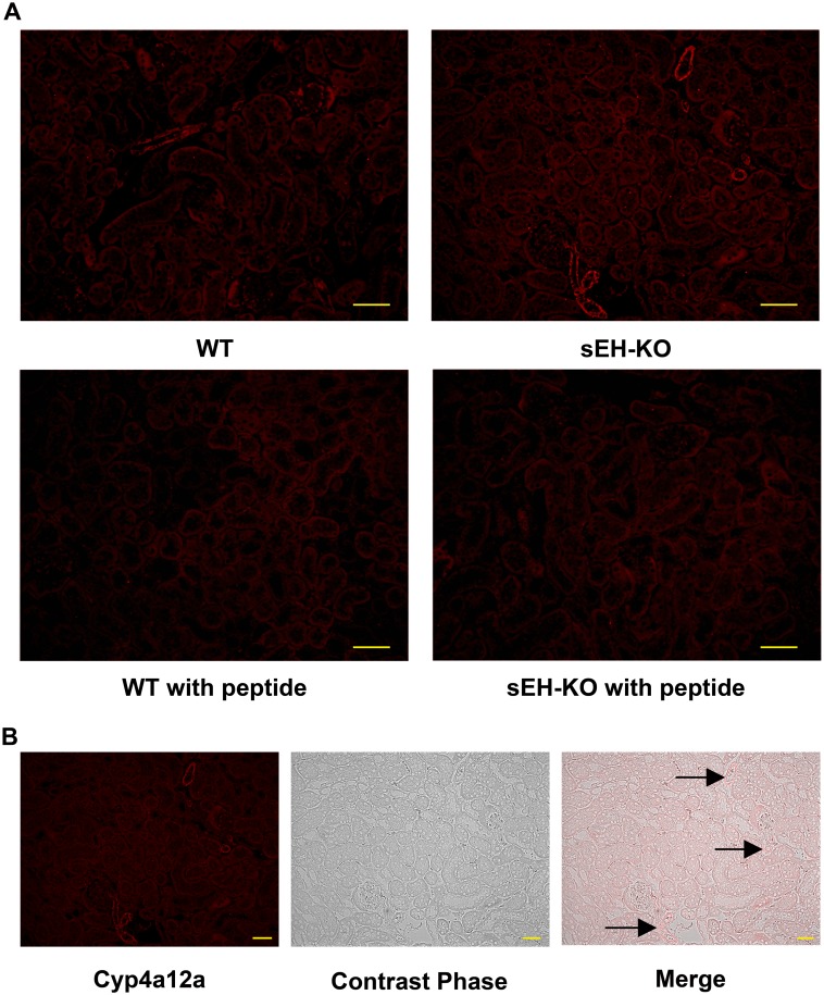
Fig 10. Intrarenal localization of Cyp4a12a protein expression.
(A): Representative images of renal sections stained for Cyp4a12a by the immunofluorescence (magnification 200×; scale bar: 50 μm). sEH gene disruption resulted in upregulating the expression of Cyp4a12a in mouse kidneys compared to WT mouse. The signals were blocked by pre-saturating the peptide-specific Cyp4a12a antibody with the corresponding synthetic peptide. (B): Images of a renal section from sEH-KO mice showing how immunostaining relates to the underlying renal structures (magnification 200×; scale bar: 50 μm). Images were taken at the area of renal cortex. Cyp4a12a immunostaining was most intense in the renal vessels (arcuate, interlobar, and interlobular arteries). Faint but specific staining occurred in tubules. No staining was detectable in glomeruli.

