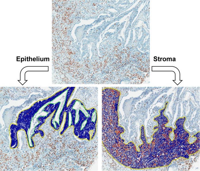
Fig 2. Immunohistochemical staining of CD68 and CD163 and measurement of the density of CD68+ and CD163+ TAMs.
Using the automatic image analysis system (ScanScope XT; Aperio) for positive pixel count v9 algorithm, the density of CD68+ or CD163+ TAM was measured separately in the epithelium (left) and stroma (right).

